Schema
Os Oppenheimer
- Fréquent
Os accessoire de l’odontoide
- Rare
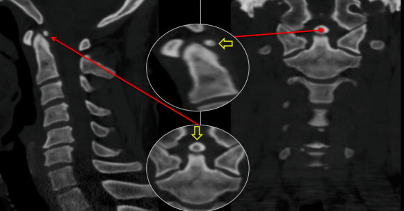
Ossiculum terminale persistant
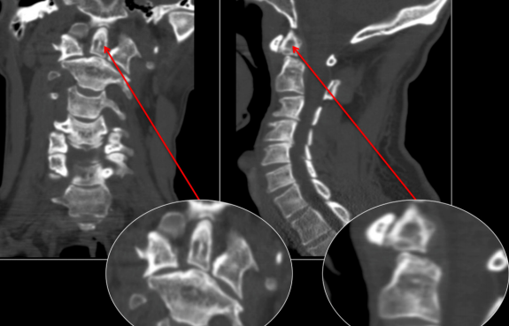
Os ondotoidum
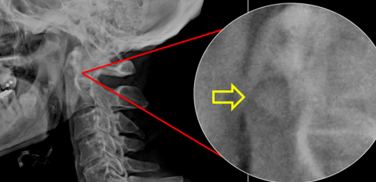
Os accessoire de l’arc antérieur de l’atlas
- Rare
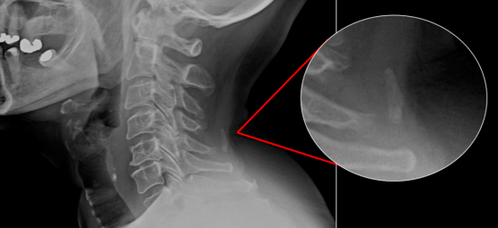
Os accessoire du ligament nuchal
- Fréquent
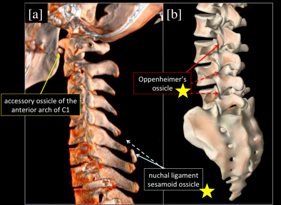
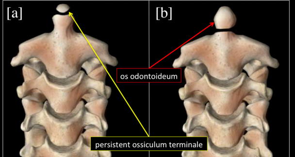
odontoid odontoide dent anterieur openheimer oppeineimer Fig. 1: 3D models of the cervical [a] and lumbar [b] spine, highlighting the typical locations of the accessory ossicle of the anterior arch of the atlas [yellow], the nuchal ligament sesamoid ossicle [light blue] and Oppenheimer’s ossicle [red] at the inferior articular process of L2. Dashed lines represent other common locations of the respective ossicles. 3D models of the cervical spine, frontal views, depicting a persistent ossiculum terminale [a] and an os odontoideum [b]accéssoires accéssoire acessoire acéssoire